Opening times
Mon 9.30am - 5.00pm
Tue 9.30am - 5.00pm
Wed 9.30am - 5.00pm
Thur Closed
Fri 2.00pm - 5.00pm
Sat 10.00am - 1.00pm
Sun 10.00am - 4.00pm
27 High St, Walthamstow, London, E17 7AD
Tel: 020 8520 4105
Email: admin@visionwise-opticians.co.uk
Retinal Detachments
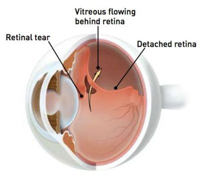
There are three types of retinal detachments. The most common form is where a break in the retina’s sensory layer causing fluid to seep underneath. This eventually causes a separation in the layers of the retina. Individuals who are particularly short sighted, with historic eye injuries or who have undergone eye surgery are most susceptible to this type of detachment. This is due to the thinner and more fragile retina in short sighted people. The second most common type is due to increased traction on the retina by strands of scar or vitreous tissue which can ultimately pull the retina loose.
The third most common type occurs when small pockets of
liquid form within a special gel (the virtreous) which usually
lines the inside of the eye. Eventually, some of this fluid moves
in between the gel and the retina, causing the vitreous to peel
away from the retina. The retina, which is like the film of a
camera, is then able to see the outer part of this gel floating
inside the eye – and this is what causes floaters.
Sometimes, when the vitreous gel comes away from the retina,
it can cause a hole or tear to appear in the retina.
This is because the vitreous gel sometimes has areas where it is
strongly attached to the retina. As the gel falls away from the
retina (a bit like wall-paper falling from the wall), the gel can
tear the retina (like the wallpaper may take a piece of paint or
plaster from the wall).
If a hole or tear develops in the retina, then there is an
increased risk of there being a retinal detachment.
A detached retina can cause loss of vision, and requires a
surgical operation to put the retina back in the right place. Thus, it is very important that you have your eye examined urgently on the onset of symptoms. There are other less common reasons for floaters – e.g. bleeding into the gel in the back of the eye from a blood vessel (usually in diabetic patients).
